How Serious Arterial Damage Connects Obesity and Stroke Risk
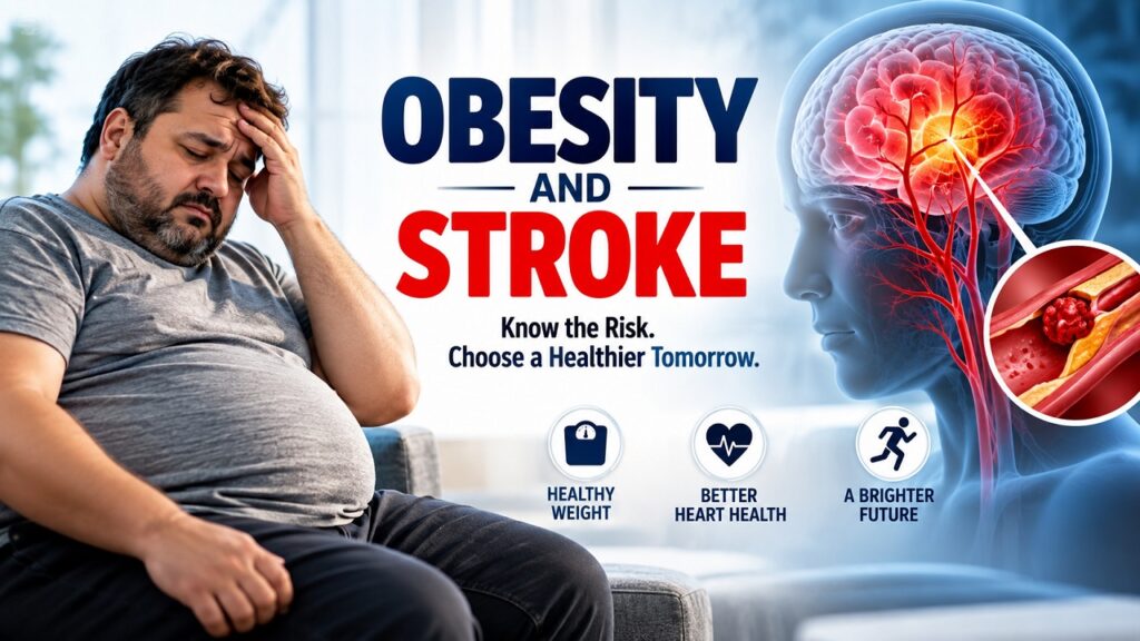
A middle-aged man sits in a brightly lit living room, clutching his forehead in pain and distress. To his right, a detailed medical illustration highlights the neurological connection between arterial blockage and brain health. The composition uses clinical blue and red tones to emphasize the serious health implications of cardiovascular strain. An educational visual detailing the link between obesity and stroke risk through anatomical diagrams and patient portraits. healthcare, wellness, stroke.
Carrying excess body weight places severe, relentless strain on the human cardiovascular system. Over time, surplus adipose tissue alters metabolism, hardens blood vessels, and disrupts regular hemodynamic flow, quietly increasing your risk of life-altering brain injury. Many people wonder whether excess weight directly harms brain circulation or if it simply operates behind secondary conditions like high blood pressure. Exploring the connection between obesity and stroke gives clarity to these concerns. This guide examines how adiposity impacts cerebrovascular function, outlines key biological pathways, and provides actionable medical steps to protect your vascular health.
TL;DR Obesity is a major, modifiable independent risk factor for cerebrovascular events, significantly raising the incidence of both ischemic and hemorrhagic strokes. Excess visceral fat actively secretes inflammatory cytokines, induces endothelial dysfunction, accelerates atherosclerosis, and creates a prothrombotic state. In addition to these direct effects, obesity drives major secondary vascular hazards, including chronic hypertension, insulin resistance, dyslipidemia, obstructive sleep apnea, and atrial fibrillation. Clinical evidence confirms that sustained weight reduction through nutrition therapy, exercise, targeted pharmacotherapy (such as GLP-1 receptor agonists), or metabolic bariatric surgery substantially lowers systemic vascular resistance and reduces stroke recurrence. Recognizing acute neurological symptoms via the BE-FAST screening protocol ensures rapid, life-saving reperfusion therapy. Comprehensive cardiovascular risk screening and personalized weight management serve as essential pillars for long-term brain health.
Quick Answer Yes, obesity is a direct and serious risk factor for stroke. Excess visceral adiposity accelerates arterial plaque accumulation, stiffens blood vessels, increases systemic inflammation, and promotes blood clots. It also triggers secondary conditions such as hypertension, type 2 diabetes, and sleep apnea. Together, these clinical changes elevate ischemic stroke risk by roughly 64% and hemorrhagic stroke risk by 24% compared to healthy baseline weights.
The relationship between obesity and stroke is an established cerebrovascular continuum in which excessive adipose tissue compromises cerebral blood flow. Obesity, clinically classified as a body mass index of 30 or higher, triggers systemic inflammation and vascular damage. A stroke occurs when arterial flow to the brain is suddenly occluded by a thrombus or interrupted by a ruptured blood vessel. When adipose-driven atherosclerosis and arterial stiffness compromise intracranial circulation, cerebral neurons become oxygen-deprived, causing rapid cellular necrosis and neurological deficits.
The Biological Connection: How Excess Adiposity Impacts the Brain
Adipose tissue is not an inert storage depot for extra calories; it is an active endocrine organ. When adipose stores expand significantly—particularly visceral fat surrounding internal organs—they release bioactive molecules called adipokines. In a state of chronic obesity, this release becomes pathological, leading to persistent vascular damage that directly threatens cerebral circulation.
Visceral Adiposity
       ↓
Pro-inflammatory Cytokines (IL-6, TNF-alpha)
       ↓
Endothelial Dysfunction & Arterial Stiffening
       ↓
Plaque Formation (Atherosclerosis) & Hypercoagulability
       ↓
Cerebrovascular Occlusion (Ischemic Stroke)
Endothelial Dysfunction and Arterial Stiffening
The endothelium is the delicate inner lining of your blood vessels responsible for regulating vascular tone, blood flow, and clotting mechanisms. In individuals with obesity, elevated levels of free fatty acids and reactive oxygen species blunt nitric oxide bioavailability. Nitric oxide is the primary molecule responsible for allowing blood vessels to dilate and accommodate changes in blood pressure.
Without sufficient nitric oxide, cerebral arteries lose their elasticity, become chronically constricted, and suffer micro-tears from mechanical shear stress. This stiffness exposes delicate cerebral vessels to high pressure, accelerating the formation of fibrous atherosclerotic plaques.
Chronic Systemic Inflammation
Enlarged adipocytes experience cellular hypoxia, triggering the sustained secretion of pro-inflammatory mediators such as:
- Interleukin-6 (IL-6)
- Tumor Necrosis Factor-alpha (TNF-alpha)
- C-Reactive Protein (CRP)
These circulating markers initiate an inflammatory cascade within the arterial walls of the carotid and cerebral arteries. Circulating monocytes migrate into the sub-endothelial space, engulf oxidized low-density lipoproteins (LDL), and transform into foam cells, establishing early-stage atheromas. Over time, these plaques can rupture, forming an acute blood clot that travels downstream to occlude cerebral vessels.
Prothrombotic State and Hypercoagulability
Obesity fundamentally shifts the balance of the coagulation system toward clot formation. Adipose tissue produces elevated concentrations of plasminogen activator inhibitor-1 (PAI-1) and fibrinogen. PAI-1 stops the body from breaking down fibrin clots naturally, while excess fibrinogen makes platelets clump together more aggressively. Consequently, when an atherosclerotic plaque tears, the body forms an abnormally dense, persistent thrombus that rapidly cuts off blood supply to vital brain tissues.
| Biological Factor | Healthy State | Obesity-Induced Alteration | Cerebrovascular Impact |
| Nitric Oxide | High bioavailability | Markedly reduced | Vasoconstriction, vessel stiffness |
| Visceral Adipokines | Balanced adiponectin | High leptin, low adiponectin | Systemic endothelial inflammation |
| PAI-1 Levels | Normal clot breakdown | Overexpressed | Impaired fibrinolysis, stubborn clots |
| Arterial Compliance | Flexible, adaptive walls | Rigid, calcified walls | Microvascular shear damage, plaque formation |
Summary: Mechanisms
Excess visceral fat secretes inflammatory adipokines and suppresses protective nitric oxide, causing arterial stiffness, chronic endothelial inflammation, and hypercoagulability. This accelerates atherosclerosis and elevates clot risks in cerebral vessels.
Ischemic vs. Hemorrhagic Stroke: How Obesity Influences Each Type
A stroke occurs in two primary forms: ischemic (caused by arterial blockage) and hemorrhagic (caused by vascular rupture). Obesity influences the pathophysiology of both conditions through distinct vascular mechanisms.
                         Types of Stroke
                               |
        -------------------------------------------------
        |                                               |
  Ischemic Stroke (~87%)                      Hemorrhagic Stroke (~13%)
        |                                               |
- Atherosclerotic Thrombosis                  - Intracerebral Hemorrhage
- Cardioembolic Occlusion                     - Subarachnoid Aneurysm Rupture
- Small Vessel Lacunar Infarcts               - Microaneurysm Breakdown
Ischemic Stroke
Ischemic strokes account for roughly 87% of all cerebrovascular events. They occur when a blood clot blocks an artery supplying blood to the brain. In patients with obesity, this typically follows three clinical presentations:
- Large Artery Atherosclerosis: Plaques accumulate within the internal carotid artery or middle cerebral artery, narrowing the lumen until flow stops or a piece of plaque breaks free.
- Cardioembolism: Obesity-related structural heart changes, such as left atrial enlargement, frequently cause atrial fibrillation. Blood pools in the fibrillating atrium, forming clots that travel straight into the brain’s circulation.
- Small Vessel Occlusion (Lacunar Strokes): Tiny penetrating arteries deep within the brain succumb to lipohyalinosis—thickening and hyalinization of vessel walls caused by persistent pressure and metabolic stress.
Hemorrhagic Stroke
Hemorrhagic strokes occur when a weakened blood vessel ruptures, bleeding directly into the brain tissue (intracerebral hemorrhage) or the subarachnoid space (subarachnoid hemorrhage). While historically linked more tightly to chronic hypertension, excess weight significantly accelerates the degradation of structural wall proteins in cerebral vessels.
The sustained physical pressure of circulating blood volume—which is elevated in individuals with obesity to meet the metabolic demands of excess tissue—leads to the formation of microscopic aneurysms (Charcot-Bouchard aneurysms) in deep cerebral arterioles. When these microaneurysms burst, the resulting hematoma exerts mechanical pressure on surrounding brain tissue, leading to rapid cell death.
| Feature | Ischemic Stroke | Hemorrhagic Stroke |
| Primary Cause | Thrombus or embolus blocking arterial lumen | Rupture of a weakened cerebral blood vessel |
| Frequency | ~87% of all strokes | ~13% of all strokes |
| Obesity Link | Atherosclerosis, dyslipidemia, atrial fibrillation | Chronic hypertension, vascular fragility |
| Onset Profile | Often preceded by TIAs; focal deficits | Sudden severe headache, rapid loss of consciousness |
| Direct Intervention | IV thrombolysis (tPA/TNK), mechanical thrombectomy | Surgical decompression, blood pressure normalization |
Summary: Stroke Typology
Ischemic strokes stem from arterial blockages driven by obesity-induced plaque buildup, blood clots, and atrial fibrillation. Hemorrhagic strokes result from vessel wall rupture caused by sustained hypertension and vessel degradation.
Does Obesity Cause Stroke? Direct vs. Indirect Pathways
When clinicians evaluate whether obesity directly causes stroke, the answer requires understanding both direct physiological damage and secondary metabolic pathways. Clinical research demonstrates that obesity functions as an independent risk factor while simultaneously worsening several downstream vascular hazards.
                         Obesity
                            |
       -------------------------------------------
       |                                         |
[Direct Pathways]                         [Indirect Pathways]
- Systemic Low-Grade Inflammation         - Chronic Hypertension
- Endothelial Shear Injury                - Type 2 Diabetes (Hyperglycemia)
- Platelet Hyperactivity                  - Obstructive Sleep Apnea (Hypoxia)
- Impaired Clot Breakdown                 - Atrial Fibrillation (Cardioembolism)
       |                                         |
       -------------------------------------------
                            ↓
               Elevated Cerebrovascular Risk
The Direct Pathway
Large-scale prospective studies confirm that individuals with elevated body mass index (BMI) face a higher risk of stroke even after researchers adjust for blood pressure, blood glucose, and cholesterol.
This direct link is driven by:
- Sustained inflammatory signaling from visceral adipocytes
- High resting cardiac output and blood volume that stress cerebral capillaries
- Chronic oxidative stress that impairs the blood-brain barrier
The Indirect Pathways: The Metabolic Multipliers
1. Chronic Hypertension
Hypertension is the single strongest modifiable risk factor for stroke, and obesity is its primary driver. Excess weight forces the kidneys to reabsorb more sodium, overactivates the renin-angiotensin-aldosterone system (RAAS), and stimulates the sympathetic nervous system. The resulting high pressure wears down cerebral vessel walls, predisposing them to both rupture and plaque buildup.
2. Type 2 Diabetes and Insulin Resistance
Insulin resistance causes persistent hyperglycemia. Excess glucose molecules bond to structural proteins in blood vessel walls through non-enzymatic glycation, forming advanced glycation end-products (AGEs). These compounds make arterial walls stiff and prone to plaque development, while also damaging small cerebral capillaries.
3. Obstructive Sleep Apnea (OSA)
Obesity leads to fat accumulation in upper airway structures, causing frequent airway collapse during sleep. Every apneic episode triggers a drop in blood oxygen (hypoxemia), surges in sympathetic blood pressure, and spikes in intracranial pressure. This nocturnal stress pattern damages blood vessels and dramatically raises the risk of waking up with an acute stroke.
4. Atrial Fibrillation
Excess adipose tissue accumulates not just around the waist, but also around the heart as epicardial fat. This epicardial fat infiltrates the atrial myocardium, creating localized inflammation and electrical disruption that can trigger atrial fibrillation. Atrial fibrillation increases the risk of an ischemic, embolic stroke fivefold.
Summary: Causality Breakdown
Obesity increases stroke risk directly through chronic vascular inflammation and hypercoagulability, and indirectly by accelerating hypertension, type 2 diabetes, sleep apnea, and atrial fibrillation.
Critical Warning Signs: The BE-FAST Protocol
When a stroke occurs, every minute matters. Roughly 1.9 million neurons die during each minute of untreated large-vessel cerebral ischemia. Patients and caregivers must recognize acute neurological warning signs instantly.
Use the clinical BE-FAST protocol to detect stroke symptoms:
- B – Balance: Sudden loss of balance, unsteadiness, dizziness, or lack of coordination.
- E – Eyes: Sudden loss of vision in one or both eyes, or sudden double vision.
- F – Face: Sudden facial droop, numbness, or an asymmetrical smile.
- A – Arms: Sudden weakness, numbness, or drift in one arm or leg when raised.
- S – Speech: Slurred speech, difficulty finding words, or inability to comprehend language.
- T – Time: Time is brain. Call emergency services immediately if any of these symptoms appear. Note the exact time the patient was last seen at baseline.
                    BE-FAST Action Flow
                             |
    ---------------------------------------------------
    |        |           |          |         |       |
 Balance   Eyes        Face        Arms    Speech   Time
Loss of    Vision      Facial      Arm     Slurred   Call
Balance    Changes     Droop     Weakness  Words   Emergency
    |        |           |          |         |       |
    ---------------------------------------------------
                             ↓
             Immediate Hospital Emergency Transport
Warning: Do not wait to see if symptoms improve. Do not drive to the hospital yourself. Emergency medical personnel begin critical neuroprotective triage, pre-notify the stroke team, and clear scanner access while in transit.
Clinical Diagnostics and Risk Stratification
Evaluating cerebrovascular risk in individuals with obesity involves more than simply checking weight on a bathroom scale. Clinicians use comprehensive anthropometric, laboratory, and vascular imaging evaluations to construct an accurate clinical profile.
Anthropometric Assessment: Beyond Basic BMI
While BMI remains the standard epidemiological metric, it has notable clinical limitations because it cannot distinguish between skeletal muscle mass and adipose tissue.
- Waist Circumference: Central adiposity correlates far more closely with vascular risk than overall mass. A waist measurement exceeding 102 cm (40 in) in men or 88 cm (35 in) in women indicates significant visceral fat deposition and elevated vascular risk.
- Waist-to-Hip Ratio (WHR): A ratio above 0.90 for men or 0.85 for women points to substantial visceral fat accumulation, signaling higher vascular risk even in individuals with a normal BMI.
Essential Diagnostic Biomarkers
A comprehensive laboratory evaluation identifies metabolic risk factors early:
- Lipid Fractionation: Assesses LDL-C, small dense LDL particles, HDL-C, and triglycerides to gauge plaque risk.
- High-Sensitivity C-Reactive Protein (hs-CRP): Quantifies low-grade systemic inflammation affecting vascular walls.
- Glycated Hemoglobin (HbA1c): Screens for insulin resistance, prediabetes, and type 2 diabetes.
- Serum Creatinine and eGFR: Assesses renal function and detects microvascular strain.
- Fasting Blood Glucose and Insulin: Evaluates the homeostatic model assessment of insulin resistance (HOMA-IR).
Advanced Vascular Imaging
When metabolic markers indicate elevated risk, physicians use targeted vascular imaging to check for arterial changes:
- Carotid Duplex Ultrasound: Measures the carotid intima-media thickness (CIMT) and identifies flow-limiting plaques in the neck’s major arteries.
- Transcranial Doppler (TCD): Measures blood flow velocity through the major intracranial vessels to identify focal narrowing.
- CT Angiography (CTA) or Magnetic Resonance Angiography (MRA): Provides high-resolution three-dimensional views of the cerebral vasculature, revealing aneurysms, dissections, or severe stenosis.
| Diagnostic Modality | Clinical Measurement | Target Threshold | Stroke Risk Implication |
| Waist Circumference | Central abdominal fat | <102 cm (M), <88 cm (W) | High visceral fat triggers systemic inflammation |
| hs-CRP | Microvascular inflammation | <1.0 mg/L | Levels >3.0 mg/L indicate higher cardiovascular risk |
| HbA1c | Glycemic control over 90 days | <5.7% | Values ≥6.5% accelerate micro- and macrovascular injury |
| Carotid Ultrasound | Intima-media thickness | Absence of discrete plaque | Stenosis >50% significantly raises embolic stroke risk |
Summary: Diagnostic Strategy
Effective stroke prevention combines waist-to-hip measurements with advanced biomarker panels (hs-CRP, HbA1c, lipids) and vascular imaging (carotid ultrasound) to identify arterial disease before an acute event occurs.
Treatment and Weight Management Strategies
Lowering stroke risk in patients with obesity requires an integrated treatment plan that combines structured lifestyle changes, evidence-based pharmacotherapy, and surgical options when appropriate.
                     Tiered Treatment Strategy
                                 |
    -----------------------------------------------------------
    |                            |                            |
[Tier 1: Foundation]    [Tier 2: Medical Therapy]    [Tier 3: Surgical Care]
- Mediterranean Diet    - GLP-1 RAs (Semaglutide)    - Sleeve Gastrectomy
- 150 min/wk Aerobics   - Dual GLP-1/GIP Agonists    - Roux-en-Y Bypass
- Targeted Resistance   - Antiplatelet Agents        - Long-Term Metabolic
- Sleep Apnea (CPAP)    - Statin / Antihypertensive    Follow-up
Tier 1: Medical Nutrition and Exercise
Cardioprotective nutrition focuses on reducing systemic inflammation rather than extreme caloric restriction:
- The Mediterranean Dietary Pattern: Rich in extra-virgin olive oil, wild-caught fatty fish, legumes, leafy greens, and whole grains. This diet reduces circulating inflammatory markers and preserves endothelial function.
- Sodium Reduction: Limiting daily intake to under 1,500–2,000 mg reduces fluid volume, easing the workload on stiffened arteries and lowering blood pressure.
- Exercise Physiology: Combining 150 minutes of moderate-intensity aerobic exercise (like brisk walking or swimming) with two weekly resistance sessions improves insulin sensitivity, promotes nitric oxide release, and lowers resting blood pressure.
Tier 2: Targeted Pharmacotherapy
When lifestyle interventions alone do not achieve sufficient weight loss or metabolic control, medical therapy can provide targeted support:
- Incretin Mimetics (GLP-1 Receptor Agonists and Dual GIP/GLP-1 Agonists): Medications like semaglutide and tirzepatide slow gastric emptying, improve insulin secretion, and reduce appetite via central nervous system receptors. Large clinical trials have demonstrated that these medications produce significant weight loss and lower the risk of major adverse cardiovascular events (MACE), including non-fatal strokes.
- Vascular Protection Medications: Antihypertensive agents (especially ACE inhibitors or ARBs) shield cerebral vessels, while statin therapy stabilizes soft atherosclerotic plaques, preventing them from rupturing.
Tier 3: Bariatric and Metabolic Surgery
For individuals with a BMI of 35 or higher (or 30 and higher with major comorbidities like severe hypertension or uncontrolled diabetes), metabolic surgery provides a durable treatment option:
- Sleeve Gastrectomy and Roux-en-Y Gastric Bypass: These procedures alter gastrointestinal anatomy and hunger-related hormone signaling.
- Clinical Impact: Long-term follow-up studies show that bariatric surgery leads to sustained weight loss, puts type 2 diabetes into remission in many patients, normalizes blood pressure, and significantly reduces the long-term incidence of ischemic stroke.
| Intervention Class | Average Weight Loss | Impact on Vascular Risk Factors | Stroke Risk Reduction |
| Lifestyle Changes | 3% to 7% total body weight | Modest improvements in blood pressure and lipids | Low to moderate (~10-15%) |
| Incretin Therapies | 15% to 22% total body weight | Major drops in HbA1c, hs-CRP, and blood pressure | Moderate to high (~20-26%) |
| Metabolic Surgery | 25% to 35% total body weight | Frequent diabetes remission; lowers blood pressure | High (>30% long-term reduction) |
Summary: Interventions
Modern stroke risk reduction combines anti-inflammatory nutrition and exercise with newer metabolic therapies like GLP-1 receptor agonists and bariatric surgery, directly addressing the underlying drivers of vascular damage.
What Most People Misunderstand About Obesity and Stroke
Public discussions on social forums, such as Reddit health communities and Quora, reveal common misconceptions about excess weight and cerebrovascular risk. Addressing these misunderstandings helps patients make informed healthcare decisions.
Misconception 1: “I am metabolically healthy, so my extra weight will not hurt me.”
Many people believe that having normal blood pressure and glucose levels completely protects them from stroke, even if they have substantial excess weight. This condition, often termed “metabolically healthy obesity,” is rarely permanent. Longitudinal studies show that over 50% of these individuals develop metabolic complications within 10 years. More importantly, excess visceral fat continues to produce low-grade systemic inflammation and mechanical strain on blood vessels, quietly advancing subclinical plaque buildup.
Misconception 2: “Stroke only happens to older adults.”
While stroke incidence rises with age, the steepest relative increase over recent decades has occurred among adults under 55. This shift corresponds with rising rates of obesity, prediabetes, and untreated hypertension in younger demographics. Strokes in younger patients frequently cause lasting disability during peak working and family years.
Misconception 3: “Only dramatic weight loss can lower stroke risk.”
Some patients believe that unless they reach a normal BMI, weight loss efforts offer little clinical value. Clinical evidence shows the opposite: losing just 5% to 10% of total body weight produces immediate vascular benefits. This modest reduction significantly lowers blood pressure, reduces circulating inflammatory cytokines, improves insulin sensitivity, and substantially reduces overall stroke risk.
        Weight Loss Benefits
                |
    --------------------------
    |                        |
5% - 10% Weight Loss    20%+ Weight Loss
    |                        |
- Lower blood pressure  - Potential diabetes remission
- Reduced inflammation  - Major plaque stabilization
- Improved insulin use  - Maximum stroke risk reduction
Common Prevention Mistakes
Avoiding key errors in prevention and care can help protect long-term cerebrovascular health:
- Relying Solely on the Bathroom Scale: Focusing exclusively on total pounds ignores changes in muscle mass versus visceral fat. A patient can lose skeletal muscle through extreme dieting while retaining inflammatory visceral adipose tissue.
- Ignoring Mild Sleep Apnea Symptoms: Chronic snoring, morning headaches, and daytime fatigue are often dismissed as simple sleep disturbances rather than potential warning signs of sleep apnea, leaving nocturnal hypertension and stroke risks unaddressed.
- Stopping Medications When Numbers Normalize: Discontinuing antihypertensive or lipid-lowering therapies once lab results improve removes the very protection that stabilized the blood vessels.
- Adopting Crash Diets: Extreme, unsustainable diets trigger lean muscle loss, lower metabolic rate, and lead to weight regain, which strains the cardiovascular system through repeated blood pressure fluctuations.
- Dismissing Transient Neurological Deficits: Passing symptoms like temporary limb numbness or brief vision loss are often written off as fatigue or pinched nerves. In reality, these may be transient ischemic attacks (TIAs) that warn of a potential major stroke.
Step-by-Step Prevention Protocol
Follow this evidence-based clinical checklist to protect your brain and vascular system:
[Month 1] Baseline Screening & Risk Profiling
    ↓
[Month 2] Metabolic & Nutritional Optimization
    ↓
[Month 3] Physical Conditioning & Sleep Evaluation
    ↓
[Month 6+] Sustained Maintenance & Re-evaluation
Step 1: Baseline Screening and Risk Profiling
- Measure waist circumference, waist-to-hip ratio, and resting blood pressure.
- Obtain a full fasting metabolic panel: lipid profile, HbA1c, and hs-CRP.
- Calculate your 10-year atherosclerotic cardiovascular disease (ASCVD) risk score with your physician.
Step 2: Metabolic and Nutritional Optimization
- Transition to an anti-inflammatory, whole-food dietary plan like the Mediterranean pattern.
- Lower sodium intake to under 2,000 mg per day.
- If indicated, consult a physician regarding modern medical therapies (e.g., GLP-1 receptor agonists) to support metabolic health and protect against vascular events.
Step 3: Physical Conditioning and Sleep Evaluation
- Start with 30 minutes of low-impact cardiovascular activity five days per week.
- Add two weekly resistance-training sessions to build and protect lean muscle mass.
- If you snore or experience unrefreshing sleep, complete an overnight polysomnography study to screen for obstructive sleep apnea.
Step 4: Sustained Maintenance and Long-Term Monitoring
- Monitor blood pressure at home weekly using an upper-arm digital monitor.
- Schedule follow-up lab work every six months to assess changes in HbA1c, lipids, and hs-CRP.
- Re-evaluate your treatment plan annually with your medical team to maintain vascular health over time.
People Also Ask
How much does obesity increase your risk of stroke?
Clinical studies show that obesity increases your overall risk of ischemic stroke by approximately 64% and hemorrhagic stroke by roughly 24% compared to individuals in a normal weight range. Even being in the overweight category (BMI 25–29.9) elevates ischemic stroke risk by 22%. Central abdominal adiposity, measured via waist circumference, serves as an especially strong predictor of cerebrovascular events.
Can losing weight reverse stroke risk caused by obesity?
Yes. Losing just 5% to 10% of your total body weight produces substantial improvements in cerebrovascular health. Intentional weight loss lowers arterial blood pressure, reduces systemic inflammation (lowering markers like hs-CRP), improves endothelial elasticity, and restores insulin sensitivity. These metabolic improvements help stabilize arterial walls and significantly decrease the likelihood of blood clots.
Why does belly fat specifically increase stroke risk?
Belly fat, or visceral adipose tissue, is biologically active and surrounds vital internal organs. Unlike subcutaneous fat under the skin, visceral fat continuously releases pro-inflammatory cytokines (such as IL-6 and TNF-alpha) and free fatty acids directly into the bloodstream. This chronic inflammation damages blood vessel linings, speeds up plaque accumulation in the carotid and cerebral arteries, and increases clot risk by elevating blood levels of fibrinogen and PAI-1.
Can young people with obesity have a stroke?
Yes. Although strokes are more common among older adults, stroke rates in young and middle-aged adults (ages 18 to 54) have risen over recent decades. This increase closely tracks the growing prevalence of early-onset obesity, type 2 diabetes, metabolic syndrome, and undetected high blood pressure. Young adults who experience a stroke face high rates of long-term disability during productive career and family-building years.
What are the earliest warning signs of an obesity-related stroke?
The earliest signs follow the BE-FAST framework: sudden loss of Balance, acute vision changes in the Eyes, asymmetric drooping of the Face, sudden weakness or numbness in an Arm or leg, and slurred or confused Speech. Even if these symptoms resolve within minutes—a presentation known as a transient ischemic attack (TIA)—they represent a critical medical emergency requiring immediate hospital evaluation.
Frequently Asked Questions
Does obesity cause stroke directly, or is it always caused by high blood pressure?
Obesity influences stroke risk through both direct and indirect biological pathways. While secondary conditions like high blood pressure, type 2 diabetes, and sleep apnea are major contributors to vascular damage, excess weight also acts as an independent risk factor. Visceral adipose tissue releases pro-inflammatory cytokines, increases oxidative stress, and elevates clotting proteins like fibrinogen and plasminogen activator inhibitor-1. These factors accelerate atherosclerosis and promote blood clot formation even when blood pressure remains within normal clinical limits. Managing weight directly protects your blood vessels beyond blood pressure control alone.
How does obstructive sleep apnea connect excess weight to stroke risk?
Obstructive sleep apnea (OSA) frequently affects individuals with central obesity due to adipose tissue deposition around the upper airway. During sleep, these relaxed tissues collapse, repeatedly stopping breathing for brief periods throughout the night. Each episode causes blood oxygen levels to drop, followed by a surge in sympathetic nervous system activity and a sharp spike in blood pressure. This repeated overnight stress damages delicate cerebral blood vessels, promotes systemic inflammation, and can trigger dangerous heart rhythms like atrial fibrillation. Over time, untreated sleep apnea substantially increases the risk of nighttime ischemic and hemorrhagic strokes.
Are new weight-loss medications like semaglutide effective at lowering stroke risk?
Yes. Modern incretin-based medications, including glucagon-like peptide-1 (GLP-1) receptor agonists and dual GIP/GLP-1 receptor agonists, have demonstrated significant cerebrovascular and cardiovascular benefits in large clinical trials. Beyond helping patients lose substantial weight, these medications improve glycemic control, lower blood pressure, reduce inflammation, and enhance blood vessel function. Major trials like the SELECT study have confirmed that these treatments significantly lower the risk of major adverse cardiovascular events (MACE), including non-fatal strokes, in high-risk patients with elevated body weight.
What is the obesity paradox in stroke recovery, and is it scientifically valid?
The “obesity paradox” refers to observational studies suggesting that patients with mild to moderate obesity sometimes experience lower short-term mortality after a stroke than normal-weight or underweight individuals. However, modern researchers view this observation with caution. Normal-weight or underweight stroke patients often have severe underlying medical conditions, such as advanced cancer, chronic infections, or severe frailty, which skew mortality comparisons. In addition, patients with obesity tend to suffer strokes at younger ages, giving them greater physiological resilience. Long-term follow-up consistently confirms that excess adiposity increases the risk of stroke recurrence, impairs functional mobility recovery, and worsens overall cardiovascular health.
The connection between excess weight and cerebrovascular disease is direct, serious, and modifiable. Excess body weight—particularly inflammatory visceral fat—places significant mechanical and chemical stress on blood vessels, leading to arterial stiffness, persistent inflammation, and accelerated plaque buildup. Left unchecked, it drives major secondary complications like hypertension, diabetes, and sleep apnea that make a stroke far more likely.
Fortunately, modern medicine provides effective tools to address these risks. Modest, sustained weight reduction, combined with targeted lifestyle changes and appropriate medical therapies, helps restore blood vessel health and protect brain circulation over the long term.
Key Takeaways
- Obesity is an independent, modifiable risk factor for both ischemic and hemorrhagic strokes.
- Visceral adiposity releases pro-inflammatory cytokines that stiffen blood vessels and promote clot formation.
- Secondary conditions like hypertension, sleep apnea, and type 2 diabetes multiply vascular risks.
- The BE-FAST protocol helps you recognize early stroke signs and access emergency medical care quickly.
- Losing just 5% to 10% of total body weight yields meaningful improvements in blood pressure and vascular health.
Actionable Next Steps
- Schedule a Metabolic and Vascular Assessment: Book an appointment with your physician to review your waist circumference, blood pressure, lipid panel, HbA1c, and hs-CRP.
- Screen for Nocturnal Symptoms: If you snore, wake gasping, or experience daytime fatigue, ask your doctor about a sleep study to evaluate for sleep apnea.
- Adopt an Anti-Inflammatory Diet: Transition to a Mediterranean-style dietary pattern focused on whole foods, healthy fats, and low sodium.
- Explore Medical Therapies If Needed: Discuss modern weight management options, such as GLP-1 receptor agonists, with your doctor if lifestyle changes alone are not enough.
- Memorize BE-FAST: Share the warning signs of stroke with your family members so everyone knows how to react quickly in an emergency.
خدمات الرعاية الطبية وإدارة السكتة الدماغية في أبوظبي
يقدم كليفلاند كلينك أبوظبي، الواقع في جزيرة المارية بأبوظبي، خدمات رعاية صحية وطبية عالمية المستوى للمرضى الذين يعانون من السمنة، وأمراض القلب والأوعية الدموية، ومخاطر السكتة الدماغية. يضم المرفق نخبة من الاستشاريين في مجالات جراحة وأمراض الأعصاب، وطب الأيض والغدد الصماء، ومراكز علاج السكتة الدماغية المعتمدة دولياً، حيث نوفر برامج شاملة ومتطورة للوقاية الأولية، والتشخيص الدقيق عبر أحدث تقنيات التصوير الوعائي، وخطط علاجية مخصصة تشمل أحدث العلاجات الدوائية وجراحات السمنة والأيض. نسعى لتمكين المرضى من حماية صحتهم الوعائية من خلال استشارات طبية متكاملة وفق أعلى المعايير العالمية. لحجز موعد أو استشارة الفريق الطبي المختص، يمكنكم زيارة الموقع الإلكتروني الرسمي عبر الرابط clevelandclinicabudhabi.ae أو الاتصال مباشرة على الرقم المجاني 80082223، كما يمكنكم زيارة موقعنا عبر خرائط جوجل (Google Maps) في جزيرة المارية، الفلاح، أبوظبي، الإمارات العربية المتحدة للحصول على رعاية متكاملة قريبة منك.